
 ENGLISH
 中文
中文 ไทย
ไทยWhy Do So Many Embryos Fail to Reach Blastocyst? Tsinghua’s Cell Study Provides the Answer
On June 11, 2026, a research team from Tsinghua University published a study in the international top-tier journal Cell. This work represents the first time that the complete process of embryo miscarriage has been visualized with clarity.
The team explicitly identified two core causes underlying embryonic developmental arrest and successfully conducted preclinical intervention trials targeting early-stage stagnation, thereby elevating the proportion of normal cells from 40% to 80%.
The First Complete View of Human Embryo Development Over 120 Hours
Many IVF families suffer embryo developmental arrest between Day 3 and Day 5, stopping embryos from growing into transfer-ready blastocysts.
Clinical statistics reveal that over half of fertilized eggs fail to reach the blastocyst stage within these five days, creating a key barrier in assisted reproductive treatment and bringing repeated disappointment to infertile families.
Human embryos take around 120 hours (5 days) to develop from fertilization to blastocyst. Traditional microscopes carry strong phototoxicity and only support 24 to 48 hours of continuous observation, leaving the full 5-day developmental process unrecorded.
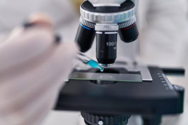
To solve this limitation, Dr. Chun So’s research team from Tsinghua University spent three years developing the world’s first high-throughput dual-view light-sheet microscope.
Equipped with low-phototoxic bilateral light-sheet scanning technology, the device takes embryo images every 12 minutes without harming samples, enabling full 120-hour embryo recording for the first time.
On June 11, 2026, the team’s findings were published in Cell. The study provided the first complete high-resolution tracking of human embryo development over the five days before implantation.
By analyzing over 150 human and cynomolgus monkey embryos and more than 2,000 cell division events, the team confirmed that early-stage (first three days) and late-stage (Day 4) developmental arrest are driven by two entirely independent mechanisms.
Finding 1: Early Arrest—Centrosome Overduplication Causes Spindle Defects
Over five days, an embryo undergoes multiple cleavage divisions. At each division, cells rely on a structure called the spindle to distribute chromosomes between the two daughter cells evenly.
Previous scientific consensus held that the first cleavage division was the most error-prone. But the Tsinghua team’s observations overturned this view.
The study found that more than 70% of embryos that arrested early had spindle abnormalities during the second cleavage division.
The team traced the root cause to centrosomes, the organelles responsible for assembling the spindle. When centrosome numbers become unbalanced, the spindle malfunctions, chromosomes are missegregated, and the embryo stops developing within three divisions.
Intervention potential: The team applied a PLK4 inhibitor (a drug that regulates centrosome replication) briefly during the second cleavage stage.
The result: the proportion of embryos with normal centrosomes increased from 40% to 80%, without any adverse effects on embryos that already had normal centrosome numbers. The team has filed international patents and is planning clinical translation.
Finding 2: Late Arrest—Endoplasmic Reticulum Stress
If early arrest is about “chromosome missegregation,” late arrest is an entirely different story.
Days 4 to 5 are the critical transition from morula to blastocyst. By conducting high-depth single-embryo proteomic analysis on embryos that successfully formed blastocysts versus those that arrested at the morula stage, the team found that late-arrest embryos had no chromosomal abnormalities.
The problem lies in the abnormal activation of the endoplasmic reticulum (ER) stress response.
The endoplasmic reticulum is the organelle responsible for protein synthesis and folding. Overactivated ER stress disrupts protein expression balance, causing the embryo to lack the key proteins needed to transform into a blastocyst, and development stops.
Intervention direction: The team is currently screening small-molecule inhibitors to address late-stage arrest. While this work is at an earlier stage, it provides a clear target for future therapeutic development.
What This Means for IVF Families
This breakthrough matters on at least three levels:
1. The reason why embryos fail to reach the blastocyst is finally visible
Previously, when embryos failed to reach blastocyst, doctors and patients could only guess why. Now, science has captured the full five-day developmental process and identified the specific root causes.
2. Early arrest has a potential treatment
The PLK4 inhibitor has already been proven in preclinical studies to increase normal embryo rates from 40% to 80%, without harming healthy embryos. Although clinical application still lies ahead, the direction is now clear.
3. Late arrest now has a target.
The discovery of ER stress involvement provides a clear direction for small-molecule inhibitor screening, and also points out the direction for the treatment of late embryonic arrest.
CEF Perspective
CEF continuously monitors cutting-edge research in assisted reproduction. The Tsinghua team's breakthrough directly addresses one of the most agonizing questions IVF families face: "Why did my embryos fail to reach blastocyst?"
Science is gradually uncovering the mysteries of early life, and providing unprecedented theoretical foundations for clinical optimization. While the journey from bench to bedside still has distance to cover, every step forward brings the possibility of higher success rates closer to reality.
If you have questions about embryo development or assisted reproductive technology, CEF can provide professional consultation and resource referral.
>For inquiries, email info@cefivf.com or visit our Contact page.